Hearing # 37 of German Corona Extra-Parliamentary Inquiry Committee (3)
This is the transcript of the Hearing No. 37 of the German Corona Extra-Parliamentary Inquiry Committee with Dr. Vanessa Schmidt-Krueger, beginning at minute 3.56.38 of the hearing to the end. The hearing was held on 30th January 2021.
RF = Dr. Reiner Fuellmich
VSK = Dr. Vanessa Schmidt-Krueger
VF = Viviane Fischer
MT = Marcel Templin
Dr. H. = Dr. Holzeisen (Italy)
Read the second part of the article
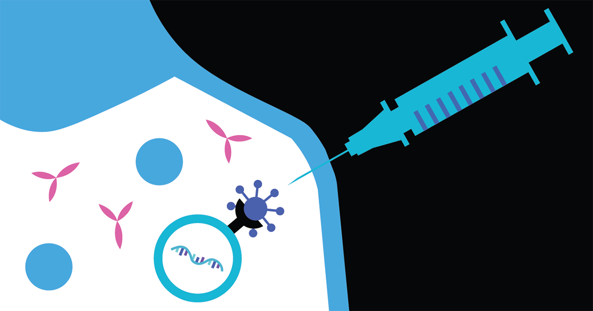
VSK: Now I’ll address the questions Ms. Fischer asked. I’ll talk about the preclinical study that BioNTech has done, largely on mice and rats. The questions that arise before something like this comes onto the market are how long it remains in the body, divided up as follows: how long do the lipids remain, how long does the mRNA remain? How are they broken down? What is their distribution in the body? The toxicology and carcinology have to be investigated. Is there a problem related to reproduction? And does it have an influence on the environment? Because we’re becoming a GMO: does this have any impact? These are fundamental questions that the EMA always has to pose.
I will refer to this Public Assessment Report – I need to say that the raw data are lacking, they aren’t in the report, which I find disappointing.
RF: Peter Doshi is challenging them on that.
VSK: Right, I’d now like to look at that data myself because I have already seen that they interpret the data incorrectly in the clinical study. I’d like to see it myself and form my own opinion. They have only provided a description of what they observed. So I can only go on that. I imagine this will be true because the observations they made have been observed by other scientists in their animal trials with their substances, too.
So what is the distribution of the lipid nanoparticles (LNPs) in the animal trial? This is similar to that described in other publications and by other scientists. They used LNPs with mRNA, not with the spike protein but with Luciferase. Luciferase has the advantage that you can make aspects visible. Useful for this trial as they they gave the lipids a radioactive marker.
If you can use the radioactivity as a marker, you can use a technique whereby can can see the organs and whether the lipids were in them or not to see. They injected the whole muscle and watched how the lipids spread out throughout the body, and found that these lipids were in many organs after just 15 minutes. Most were at the injection site, in this case it was the muscle, but a lot in the plasma, too. Logical because it’s transported in the plasma, but also 22% in the liver. And if you inject it into the veins then 60% of the cationic lipids can be found in the liver, and 20% of the PEG lipids. They were also found in the spleen, the adrenals, and in both sexual organs. Further organs were not described. So I assume that it spread out throughout all organs. It is basically absorbed everywhere where blood flows. The description focuses most on the injection site, the plasma, and the liver.
Then they looked at how the lipids were degraded. They found evidence of the cationic lipid in the plasma for 12 days, and evidence of the PEG lipid for 6 days. So they remained for quite some time. There isn’t any more information, so I don’t know whether the lipids could be evidenced for longer or not. 50% of the PEG is degraded via excretion, i.e., it is excreted from the body. It goes into our “sewer system”, as it were. The cationic lipids are exclusively degraded in the cells, only 1% was found in the stool. This means the cells take the full hit of the toxicity. Then they analysed the half life of this cationic lipid in the liver, they say it is 3 weeks. With half life at the beginning the substance always degrades faster, and then it gets less, the curve gets flatter. This half life at the outset is already 3 weeks, which is relatively long. And how long does the elimination take? One can still find 5% of the lipid in the liver after 4 – 6 weeks – that is incredibly long, and with the PEG the half life is 1 week. So it is shorter, but because a large proportion, i.e., 50%, is excreted. That is not the case with the cationic lipid.
We don’t have any other information or investigations regarding other organs, they just investigated liver, plasma, urine and stool. They should definitely have looked at other organs. Perhaps they did, but there’s nothing in the publication about that.
And then they looked at how fast the RNA is degraded. This is where the Luciferase comes into its own. The Luciferase can transform a substrate so that one sees it in colour, it fluoresces. You can detect it. But it’s not a very sensitive method. And they only injected 2 micrograms of RNA. 30 micrograms are being used for us. This means what you are seeing is probably a lot stronger in the case of the actual vaccine being used. So in the muscle where it was injected there was a peak after 6 hours. First the LNPs have to be taken up into the cell, the protein has to be formed, this Luciferase, and only then does the reaction take place. You see this after a max. of 6 hours, it is taken up by the cells extremely fast, and the protein is also expressed very fast. You can still see the protein after 9 days. There are publications – there is one from 2016 for example – where they say that one can see the Luciferase for 35 days, but that always depends on how stable the RNA is, and they didn’t do it with the spike RNA but just with the luminescence, and the spike RNA may very well have a different stability. So they didn’t investigate it properly for our vaccine, I would say.
In the liver they saw a peak after 6 hours, and after two days it was gone. This is because the liver has a very high metabolic rate.
So to summarise, both the RNA and the LNP are taken up relatively fast. And the cationic lipids remain in our bodies for a very long time. This was also interesting. There seems to have been a discussion of the EMA with BioNTech about the period that it remains in the body: how long is it in the case of human beings, they asked, because the study wasn’t done. BioNTech referred to a study from 2010, by Mamoth et al. I have not been able to find this in the publications database, and there is no list of references below the EMA report, so I don’t know whether this is true at all and whether that article exists, but they say they have used similar lipids, and when they calculate the conversion from this mouse or rat study to human beings, that cationic lipids have a half life of 20 to 30 days in human beings, and the elimination to 5%, so not really eliminated, takes 4 – 5 months. They assume 4 – 5 months, and the EMA Committee just said “That’s a long time”.
Dr. H.: The second vaccination comes on top of that after 30 days …
VSK: Yes exactly: none of that has been investigated. Basically they haven’t conducted any kinetics with this vaccine. Not on the mice either. The LNPs were the same [… Inaudible], but the RNA was different. They should really have done it with the actual vaccine. They should have marked it and then carried out the whole study again. They didn’t do that.
MT: I’d just like to ask a question. You said something was excreted from the body. Is there any danger that people who are vaccinated could be causing as a result of this, or is it excreted and then it’s gone?
VSK: That wasn’t investigated.
Dr. H: Oh God.
VSK: There’s no data on that.
MT: I reckon we’ll need to be drinking spring water from bottles from now on then …
RF: That doesn’t sound good. And what kind of consequences does that have? You’ve got apoptosis that apparently takes place throughout the body, as you have just told us: what does that lead to?
VSK: Yes, I can tell you that in a moment, that’s the toughest of all to hear. But I just wanted to finish talking about the elimination, they haven’t considered this at all because they haven’t done any analyses on the environmental impact of all of this – as I said, we’ve become GMOs, it is possible that modified cells are eliminated: think about the lipids, the RNA from the vaccine – We know that the lipids – the PEG at least – are being excreted. What occurs to the sewage if so much is being eliminated? If so many lipids are in it? Does this cause a problem, or is it degraded? We just don’t know that. I don’t know, I’m not an expert in how it is degraded.
Dr. H: Exactly, and intentionally one has to say as a lawyer, in July 2020 EU legislation was changed: EU legislation on GMOs was declared inapplicable to the vaccines. That’s when this monstrosity began, from a legal perspective. We will be addressing this with a plea for annulment. This is opening up a horrendous abyss – unbelievable.
VF: This stuff, when it appears in the sewage works, and if it were not filtered out – If I drank this stuff from the laboratory: would that cause a problem? If I drink these lipids? Are there investigations on that, if it gets into the water supply, or I drink a little from a glass, what would happen?
VSK: I can only say that if we excrete the PEGylated lipid and if it is not filtered out and one drank it, i.e., assuming it doesn’t get degraded naturally in some other way, it would continue to exist in the drinking water (and I don’t know that for certain), then you would have a problem if you had an allergy to it.
That would be the same as these anaphylactic shocks.
I could imagine that. It is absorbed via the stomach, but I don’t know exactly what happens then – whether there would be allergic reactions. It’s possible that it is completely degraded in the stomach by the enzymes that break down fat.
MT: I find it fascinating: we are talking about gene technology, there are people who have been warning for years and saying if I eat gene-manipulated corn or make bread out of it, then I could grow a second head, maybe 7 heads – I’m astonished that people aren’t looking at this more closely and that it’s the same people who are keeping their mouths shut. The same ones who before said this is so terrible, we can’t do that.
RF: If doctors don’t learn to think about what’s really occurring, and just vaccinate away instead of listening to people like you, just blindly trusting and not asking any questions …
But just to go back a moment: If this spreads out in the body, to all the cells, then that’s a horrendous scenario if you say the result is that massive numbers of cells self destruct.
VSK: Yes, that’s a good introduction to that exact point, with all the consequences. Let’s talk about the preclinical study – about what occurred to the rat.
In the preliminary experiment the rat was injected in the muscle with 30 micrograms of this same vaccine that is now being used. That is comparable, but three times rather than twice. At intervals of one week. And two days after the last injection, that would have been 17 days after the first, an autopsy was conducted, and the following was found. As mentioned, I don’t have any raw data, only descriptive written data. The rats had an immune response, raised lymph nodes, the spleen, cell numbers, that is all normal, increased production of lymphocytes, i.e. B and T cells in the bone marrow, production of neutralising antibodies, circulating white blood cells, cytokine release, that is all normal.
But then other aspects followed: Their body temperature was 1 degree raised, that is also normal, a slight temperature, for rats too;\ but their body weight went down although they were having their normal feed … With rodents, if the body weight decreases, that is always a sign of massive stress. And then they did an autopsy. They document damage to the muscle. What they make public – swelling, oedema, reddening – is just the tip of the iceberg. I’ll dissect this in a moment for those who are unfamiliar with the specialist terminology: myofascial degeneration, scleropathy, encrustation accompanied by spread of this inflammation to adjacent tissue, subcutaneous inflammation, hyperplasia. So what does all that mean? Subcutaneous inflammation means the lowest layer of skin – the skin has three layers, and the bottom one is inflamed, that is the layer where the adipose cells, nerves and blood vessels are located. If these become inflamed then the adipose cells burst open, the fatty acids are released, and further accentuate the inflammation.
This results in scleropathy, i.e., the tissue hardens because increasing amounts of connective tissue are formed. This is ultimately like scar formation. The tissue is so heavily damaged …. If you cut your finger and it is superficial then the upper skin layer can regenerate, you don’t see anything afterwards. If you cut yourself too deeply and it goes through all three layers of skin then the organism can’t replicate its own structure. Then, because the cells need to be replaced, the wound is necessary to be closed, connective tissue is formed, deposits, a scar develops. And this is the case with the muscle, it hardens due to the deposition of connective tissue. This is called fibrosis. The tissue basically loses its function at these locations, encrustations develop, this is the deposition of salts in necrotic tissue; necrotic tissue is tissue that is dying. The muscles there are dying. They talk about myofascial degeneration, this means death of the cells of the muscle fibres, which is simply replaced by connective tissue that is non-functional.
VF: Is that just local or at many locations?
VSK: At that location it’s only local, only in the muscle. You can see this entire process from the blood parameters that were measured: they noted for example a 72% increase in alpha-2-macroglobulin, this means the increase is part of the immune response due to inflammation, but you also get an increase in alpha-1-acylmycoprotein […], that is formed when there is a particularly strong injury in the tissues, caused by inflammation or infection, in this case from the vaccination, and an increase in fibrinogen…. That is a sign, when that is elevated in the blood, of inflammation of the blood vessels, it is basically responsible for blood coagulation. I have said that the blood vessels are in the bottom layer – the blood vessels are damaged, and this is probably why fibrinogen is formed, to reseal the blood vessels. I wonder, with the elderly in care homes, they are often on anti-clotting medication as a prophylaxis: is it possible that their coagulation doesn’t work properly? – You need coagulation: maybe it doesn’t function correctly? Can this have consequences if the blood vessels are heavily damaged due to this vaccination?
RF: We will see all of that very quickly, I fear.
Dr. H: The side effects, i.e. the correlation with other medications, was not examined at all. This can expressly be seen from the appendices to the EU Implementation Decision for both vaccines. I find what you are now telling us absolutely criminal.
 VSK: So that’s what occurs locally, at the site of the muscle. We have heard that a great deal goes to the liver, and that is a bit more serious. This leads to hepatocellular periportal vacuolisation. On the day of the autopsy, where they found it, and probably a lot earlier, because it gets into the liver relatively fast and then that takes place relatively quickly. So what does that mean? Hepatocellular means relating to the cells of the liver. Periportal means the liver cells near the portal vein. That is the place where the blood enters the liver. I.e., this damage will not be caused by anything else in the rat. If the rats drank alcohol, ok, then this damage would also occur, but it would be across the entire liver. But this is something which is entering via the blood flow, and only in the proximity of this vein, and there one particularly sees the damage. And they are so damaged that they are vacuolising: that is always an indication that the liver cells are dying. I’m loathe to use the word poisons, but the liver is trying to sequester away the substance that is damaging to it; it doesn’t manage, and the the cationic lipids are the perpetrator, BioNTech admits that themselves, that’s in the report, it’s the cationic lipids. The liver tries to eliminate these cationic lipids, to metabolise them, but doesn’t manage because there are too many of them. The volume is too great. So it tries to ferret them away in an area of the cell, and that is when vacuoles arise in the cell: water streams in, it’s simply an area where they no longer do any harm. But when these vacuoles arise then the function of the liver cell is massively disrupted, many of them die, they lose their function. They self-destruct, commit apoptosis. So that’s what occurs in the liver.
RF: If this is found during the autopsy then it seems to me to be a clear indication that it has been caused by the vaccination. Or can there be other causes for it? You have just said that if you drink alcohol this occurs too, but not like that. What is found there seems to be a sure sign that it was the vaccine that led to the death.
VSK: Yes, one can also investigate what there exactly is in these vacuoles. One can look and see whether the cationic lipids are there. If you have a vacuole caused by alcohol, you have a fatty liver; the alcohol is made into fat, it is stored in fat. That’s this steatosis that one hears about.
RF: I just wanted to ask – independently of the severe medical malpractice that was just described previously: you can find out what the cause is via an autopsy can’t you?
VSK: Yes, you can I’d say. And you can also see that the liver is severely damaged from the blood parameters, doctors should know this really. These are standard values: an elevation in GGT, an enzyme, can have various causes. It’s definitely an indication of liver damage from medications or poison, for example. It is an indication that the liver cells are dying, that is when increased GGT is secreted.
And then we have elevated AST. This is a metabolic enzyme that goes up in liver inflammation and cardiac damage. There’s elevated alkaline phosphotase – this is produced by the bones and liver, for example, and one or two other organs; an increase points to liver and bone injury. And then we have a decrease in the ratio of albumin to globulin. This ratio is always measured to see whether the volume of protein in blood is constant. If not, it is a sign of disease: too much protein is being eliminated. If a decline is noticed, this is a sign of severe liver damage, inflammation, a digestive disorder, etc. The rat is displaying a loss of protein.
To summarise, one can say that the liver is massively damaged, and the liver cells are dying.
They did say that after the autopsy, three weeks later, the liver had regenerated. But the EMA didn’t discuss what the situation might be with people who have a liver disorder, who don’t have this regenerative capacity. What of those who have hepatitis or an alcoholic liver or whatever? Who had been living an unhealthy lifestyle? If something comes on top of that, you can very quickly get organ failure. This shouldn’t be forgotten, it needs to be discussed, but it’s being completely swept under the carpet.
So why exactly is the liver being damaged? It’s because the liver is the organ that takes up the most lipoproteins. And why does it take up the most? Because one of its functions is to break down cholesterol; I’ve explained that the nanoparticles are bound to ApoE proteins. These make their way directly back to the liver where the cholesterol is broken down, and that’s why the liver comes into contact with a huge amount of this.
RF: I just have to reiterate: how can they be vaccinating against this backdrop?
VSK: That’s not the whole story. You get inflammation of the perineural tissue of the iscias nerve, the strongest nerve in the body. And then inflammation in the extracapsular tissue was found, I don’t know exactly what capsules they mean, they didn’t specify that, but I assume that’s the joint capsules. What about people with arthritis for example? And then this is particularly important, very dangerous: they found a moderate to strong reduction in red blood cells and reticulocytes in the bloodcount. That accounts for the hypoxia. They are massively damaged by the lipid nanoparticles. Why is that? Because it is exactly these red blood cells that are used as a cell model for oxidative stress, they are particularly sensitive to oxidative stress. Because they carry the haemoglobin. All cells that carry oxygen are always sensitive to oxidative stress. And when the LNPs get into them and cause this massive oxidative stress, they die very quickly. So the rats would have to be suffering from hypoxia or at least they found that they had less haemoglobin because obviously that is gone when the cell is gone, and lower haematocrit. These are very clear signs of hypoxia, and I have to say this needs to be looked at very critically, because what about people with cardiac disorders for example. A cardiac muscle, for instance, if it is undersupplied with oxygen, this can very quickly turn into a heart attack. And as far as I know there is someone who had a heart attack after vaccination. I’m not saying that person died of it, but one should at least look into it.
RF: You don’t need to say that, we don’t either, because based on what you have already described earlier – they’re using triple the volume they need to for example – we will find various forms of serious medical malpractice. The doctor is responsible and will need to prove that this did not occur due to the vaccine and that he/she is not guilty, or at least negligent in what he/she has done. They’re not going to get away with it.
Dr. H: Exactly.
Read the fourth part of the article
yogaesoteric
September 9, 2021