Our hearts have consciousness and are connected to our entire body
Traditionally, the heart is viewed as just being a pump that propels blood through your body. However, as we will discuss in this article, many aspects challenge that assumption.
For example, tubes of liquid crystalline water exposed to infrared energy (which is everywhere) will spontaneously create fluid flows within them. Since the blood vessels are designed to do this, it’s entirely possible that liquid crystalline water may be the driving force behind fluid circulation in the body and the heart simply functions to convert this continuous flow into a pulsatile wave.
Likewise, organ transplants profoundly undermine our current conceptions of reality as they have demonstrated that much of what we consider to comprise our “consciousness” in fact originates from the heart, not the brain as memories, talents and preferences from a donor are observed to transfer to the organ recipient. This suggests that the heart has an immense degree of innate intelligence and in this article, we will explore how its intelligence makes life possible.
Conjugated Heart Ties
Years ago, I came across an intriguing paper by a team of Russian physiologists led by Dr. Goncharenko. It took me years, but I was eventually able to find a colleague who knew the researchers and received a copy of their research. Since most of it does not exist online, I posted a second article containing it. Everything henceforth will reference that compilation.
Note: Before we go further, I want to note that I feel very strongly about the immorality of animal testing, as many lab animals demonstrate clear signs of being conscious and are regularly horrifically tortured to death for often completely unnecessary experiments. Furthermore, I think we all pay a price for this cruel experimenting because it creates a system of medicine where a similar tendency is often directed towards human beings, leading to a variety of horrific abuses, like the forced sterilisation initiatives that have been conducted throughout history such as the hCG vaccination campaigns.
I share this because I am still quite on the fence about how I feel about the research I will cover in this article – what the researchers discovered was amazing, but those discoveries required very cruel acts to be done to animals.
Dr. Goncharenko’s research was originated from a study in the 1970s where six baboons were subjected to a very mean situation in order to see if the stress of it could cause a heart attack. The final baboon had the greatest stress response and eventually had a heart attack. When an autopsy was conducted, it was observed that a fatal heart attack had occurred in a very specific site in the heart that was accompanied by the typical thrombus [clot] seen at the site of a heart attack. However, a curious observation was also made. A large hematoma was found in the left iliac artery (suggesting damage had occurred to the artery during the experiment), and at that arterial hematoma, six thrombi were found matching the thrombi in the heart. Since no other thrombi were found in the arterial system, this suggested the heart was inexplicably directing thrombi from it to the site of the injury to repair it.
While investigating this, the researchers recalled another curious observation repeatedly made throughout the history of medicine; that blood in different blood vessels differed in its composition. For example, blood to the brain is warmer and contains younger red blood cells (which are better able to nourish and meet the needs of the brain), something also seen when an actively exercising arm (which needs the healthiest blood) is compared to a resting arm (this has also been found when comparing an exercising hand to a broken one).
Conversely, blood to the spleen (which breaks down blood cells that have aged and lost their viability) typically receives the older and weaker blood cells (although in cases of carbon monoxide poisoning, the heart somehow knows how to avoid shunting weakened blood cells to the spleen for half an hour). Other examples occur as well, for instance, the blood that goes to a pregnant woman’s uterus has more nutrients than the blood the rest of her organs receive.
Note: One of my mentors (whose judgement I trust) believes the spleen not only destroys the non-viable blood cells it receives but also imparts a vitality to the blood cells (this used to be believed by earlier American schools of holistic medicine and is a foundational belief of Chinese medicine) and restores the weakened blood cells so they can continue to function. My mentor in turn argued this may explain why the splenic artery is one of the largest arteries in the digestive system. This proposed function is also somewhat congruent with the observations in many holistic disciplines (e.g., Chinese Medicine) that fatigue is associated with a dysfunctional spleen. Additionally, the mRNA vaccines are known to accumulate in the spleen, and I have met a few people who had splenic infarctions (blood clots) after the vaccine, so I have wondered if this may partly explain the fatigue frequently observed in the vaccine injured.
With their preliminary data, the researchers decided to repeat the initial experiment and discovered that for monkeys, dogs, rats and rabbits the same phenomenon was observed. If a specific artery was injured, multiple conchoidal (spiral) shaped thrombi that contained heart tissue would appear at the site of injury and nowhere else created a layered clot. Reciprocally, a specific part of the heart would experience a myocardial infarction (heart attack) when this occurred and the conjugation between the specific artery and part of the heart, was similar in all the animals and identical for animals of the same species. This suggested the heart was sacrificing part of itself to protect a blood vessel from developing a critical injury.
Conversely, they also found the reverse was true. Injuring a part of the heart would cause an injury in its conjugated (paired) artery 1-2 weeks later. For example, a trauma to the upper heart of a rat caused tail necrosis, while doing something similar to rabbits and dogs caused a gradual atrophy of their hind limb muscles. This suggests that each part of the body has a part of the heart responsible for getting blood to it, and if that part of the heart stopped working, the paired area would gradually weaken from a loss of blood flow.
With more work, they determined the part of the heart that mattered was on the inner lining of the heart (the endocardium), so damaging the heart’s surface but not going deep into it did not create a reciprocal injury in the vasculature. Likewise, while the location of the endocardium lacerating an artery damaged stayed consistent, the region of the myocardium affected by that laceration had some variability.
The specific areas that were damaged in the heart from a peripheral injury were the Tebesian vessels (discussed below) located within the left ventricle, the right ventricle and the left auricle.
Note: Goncharenko uses a few differing spellings for these vessels in his writings, which I assumed was due to a translation issue.
To further validate the heart conjugation, they experimented with putting a radioactive tracer (albumin with iodine-131) into the parts of the heart conjugated with the lower limbs and found a brief spike of radiation was seen in the conjugated artery in the lower limbs which was many times greater than the radioactive spike throughout the rest of the body (approximately 10-fold). Next, they tried repeating the experiment, but this time destroyed the Thebesian veins and the trabeculae carneae in the conjugated region of the heart prior to injecting the tracer into the area. Once this happened, there was no longer a radiation spike in the lower limbs (the radioactive concentration was equal to or lower than that observed in the other sites monitored throughout the body).
While it was not ethically possible to repeat these experiments in humans, there were a variety of observations that suggested the same thing was occurring in our species. Certain surgeries (e.g., a gastric or duodenal resection, nephrectomy, hysterectomy, or thoracotomy) require ligating an artery (cutting off its blood flow), and there are numerous reports of beings having heart attacks during those surgeries. Goncharenko’s team was able to do autopsies on some of those cases and discovered the same thrombi clustering at the site of the ligation he’d seen in the other animals. Likewise, there are thousands of reports in the medical literature of an arterial injury causing a heart attack. From these reports and his own observations, he compiled numerous cases where both the specific injury site and heart attack site were known, such as:
- A stab wound to the left femoral artery caused a heart attack at the front apex of the heart wall.
- Crushing injuries of soft tissues of the left hand finger led to a myocardial infarction in the anterior-lateral wall of the left ventricle and septum.
- A fracture of the left radius with bruises led to a patchy infarction on the back wall of the left ventricle.
- Operations in the carotid arteries can lead to the infarction of the front wall of a heart base and the interventricular septum.
- Operations on a hepatic artery lead to the damage of the front and middle portion of the side wall of the left ventricle.
- Renal damage leads to a myocardial infarction of the anterior wall.
- Single gastric bleeding in peptic ulcer disease led to the emergence of an infarct in the rear-side, rear and rear-septal region of the left ventricle of the heart.
- Bleeding from the mesenteric artery was accompanied by a massive heart attack in the front-septal and apical regions of the lateral and posterior wall of the left ventricle.
Conversely, he noted that operations on the cardiac base (which conjugates blood flow to the brain) were known to create disorders suggestive of impaired blood flow to the brain – such as encephalopathy, mental depression, changes in behaviour or intellect, epilepsy, stroke and visual impairments. Likewise, heart attacks in specific areas of the heart have been known to cause necrosis of the nose, ears, arms and impotence.
Note: Similar brain damage also occurs when a patient is put on a heart-lung machine (e.g., during a heart surgery) which suggests something besides just pumping blood to the brain is needed for its health.
Lastly, rheograms have sometimes been able to monitor arterial flow at the same time heart damage occurred (either spontaneously or during heart surgery) and have shown that blood flow dropped off in a specific artery at the same time heart damage occurred.
The conjugations they found were described as a “Phaistos spiral disk with pictures of acupuncture of the ear, palms or soles of the feet” and depicted as follows for mice and humans:
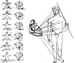
Goncharenko also provided the following descriptions for those conjugations:
- The projections of these areas on the heart of all animals were of the same type, but their sizes varied.
- For example, the inner surface of the apex of the left ventricle is associated with the vessels of the left hind limb, and the area to the right and back to the tip – with vessels of the right hind limb.
- The middle part of the ventricles, including the septum of the heart, occupies projections conjugated with vessels of the liver, kidneys, and the surface of its rear part is related to vessels of a stomach and a spleen.
- A surface located above the middle of the outside of the left ventricular cavity is a projection of vessels of the left forelimb; the front part of the transition to the interventricular septum is a projection of lungs, and the surface of the base of the heart has a projection of brain vessels, etc.
Additionally, Goncharenko also found heart conjugations for the adrenal glands and the thyroid.
Note: Over the years, I’ve asked bodyworkers who treat the vascular system and the heart (they are hard to find) about these conjugations. The most talented ones have told me they can both consistently feel a connection between regions of the heart and specific parts of the arterial system along with an energetic connection between different parts of an artery (e.g., the left or right of the abdominal aorta) and where that blood will eventually end up in the body.
Based on their answers and Goncharenko’s research, I am inclined to believe the heart can tell when an artery has been damaged or an organ needs help, but I am at a loss to explain how it is able to do that. The researchers tried to assess the most obvious mechanism (signals from the nervous system) by using an intralumbal or local procaine block and found that anesthetising the nerves for the injured artery had no effect on the heart’s ability to detect the injury, which indicates something besides the nerves gave the heart that awareness. Later they also tried disabling the central nervous system of mice through the shock of blood loss and while this altered the clotting process, it did not disable the heart’s conjugating ability either.
Goncharenko also shared that the heart conjugation may be a natural evolutionary process:
Wormlike animals have hearts in every one of their segments. There may be several dozen of them. With an increase of the body complexity this number fits in four hearts and in mammals – in one. But although many hearts united in one, they continue to provide blood to organs once related to them.
Lastly, Goncharenko posited that there might be a “heart” for the heart which acted towards the heart as the heart did towards the entire body (e.g., by sorting and directing blood). He concluded the auricles (now known as the atrial appendages) were the “heart’s heart” – for example, infarctions (heart attacks) occur in the auricles and blood clots from those auricles can be found within the coronary arteries. Since Goncharenko believed the auricles could be the source of mysterious sudden deaths, I’ve thought quite a bit about how they relate to the wave of sudden deaths we are seeing now.
However, while it was possible to prove this conjugation was occurring, it also posed a much greater question: How was it occuring?
Spiralling Motion
I encountered Victor Schauberger’s research which concluded that the ideal way for water to travel (both so it was energised and so it had the minimal amount of resistance) was in a spiralling vortex where everything carried within the water was concentrated in its centre. Schauberger’s conclusion was heavily influenced by his observation that streams and rivers would adopt curved patterns (both horizontally and on the bottom of the riverbed).
This suggested water was trying to flow in a curved motion (along with gradually moulding the waterway to match its vortexing motion), and that the consistency of this pattern indicated it was the most energetically favourable way for water to travel. Furthermore, he found with rivers losing their riverbanks (which is a significant issue in certain areas), the best way to fix it was not to continually reinforce the riverbank, but rather to place boulders in the bottom of the river that restored its vortexing motion. This specifically worked because it reduced the amount of friction on the edges of the river (as the force of the water’s flow and the solid objects within became concentrated at the centre of the body of water). Assuming Schauberger’s observations are in fact true, it would suggest that natural selection would favour a similar architecture in biology. The benefits of reducing the energy needed to move blood through the body, and more importantly to reduce the damage blood flow causes to the lining of the blood vessels (which I and others believe to be the primary cause of heart disease) are both critical for an organism’s survival.
If blood were to travel as a spiralling vortex through the arteries, two elements would be necessary:
- Something to initiate the spiralling motion.
- Blood vessels with a curved shape that create the vortex (a manner not all that different from what Schauberger designed for rivers).
As it so occurs, there is such a curved shape to the arteries throughout the body, something I have seen best demonstrated by plasticised cadavers (e.g., see this video, or this high-resolution image of a heart, a structure which is also curved to facilitate the spiralling motion of blood). Additionally, a few vascular workers I’ve asked about this have shared that there is a very clear spiralling pattern they can feel on the inside of the arteries (they specifically notice this because sometimes it gets knotted up) – which if true, suggests a similar architecture to the systems Schauberger designed creates a spiral of anything flowing through the vessels.
Heart Vortexes
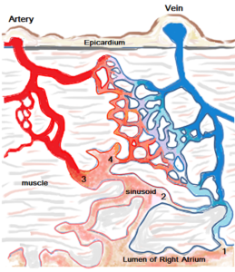
Goncharenko’s team eventually figured out a fairly unknown part of the heart, the Thebesian vessels were the responsible party. Roughly 200 of these tiny veins (first discovered in 1706) line the inner surface of the heart (with approximately 100 being in the left ventricle) and drain blood from the heart’s inner chambers. Many questions still exist regarding these veins:
Through Thebesian vessels and trabecular cells per day passes 40% of coronary blood – almost 700 litres. This “useless” circulation of large volumes of blood in different directions, not feeding the myocardium, still is not understood. When pouring plaster cast in the left ventricle the excretory channels become visible. They spiral from the apex to the base, along them there are dozens of mini-hearts, the location of which is reminiscent of the primitive worm curled in the heart.
The first hint the Thebesian vessels were responsible was that in the affected area of the heart, there would be destruction of some of the trabeculae carne (tiny muscular columns that line the inside of the heart) and thrombi (along with tiny haemorrhages) forming at the Thebesian veins. Conversely, as mentioned before, these are the same vessels which when destroyed in the heart stopped conjugation from continuing to occur.
The second came from the work of another anatomist who filmed the Thebesian vessels spouting vortex-shaped microjets during diastole (when the heart fills with blood).
The third came from the Thebesian vessels orifices having valves and opening into the trabeculae, leading Goncharenko to suspect the Thebesian vessels were using the trabeucalae to sort the blood they took in. This led Goncharenko’s team to refer to the trabeucalae as “mini hearts” due to their ability to function independently of the rest of the heart.
The fourth was knowing that the vortex structures are the most stable form of liquid movement (they keep their form for a long time) and thus were the ideal way for the heart to sort blood flows directed to different areas. Later they were able to create an artificial model that showed vortex flows they made could be directed to a target location.
Note: To further support this theory, Goncharenko cited that the primitive lungfish, which is able to separate arterial and venous blood within the same ventricle, and then direct the venous (deoxygenated) blood to its gills and the arterial blood to the brain – something that requires a heart chamber to both divide blood into separate types and direct its flow with vortices. Additionally, he also argued the same process occurs in the human foetus before the complete blood vessel network forms.
Lastly, they concluded each of the individual micro-vortex would merge to create a combined vortex that exited the heart before separating into the individual vortexes that travelled to their specified parts of the body. This again mirrors the vascular workers’ experience that once blood leaves the heart, it feels as though exactly where it will end up in the body has already been decided. To test these theories, they inserted Evans blue dye into a rat’s heart during systole (the contraction phase) and flash-froze the rat (with liquid nitrogen) and then examined slices of the inside of its heart under a microscope. From piecing together these two-dimensional images, they found that vortex-like conglomerates of red blood cells formed on the transit arteries that were lined with villi (microscopic fingers) which merged to become the Thebesian vessels. The conglomerates, in turn, were toroidal (doughnut) in shape, always contained micro-bubbles in them, and I believe (based on the wording of the paper) often had other blood elements in the centre of the red blood cell toroid. In later writing, Goncharenko referred to these conglomerates as resembling spindle architectonics.
Next, they switched to moving injecting the dye 5-8 times every 2-4 heartbeats, then flash-freezing the mice, cutting their vessels into thin sections, and then looking at each two-dimensional image to reconstruct into a three-dimensional model of the blood flow. They discovered that the toroidal blood structures would transform into vortexes as they left the heart that were maintained by toroidal rings of red blood cells surrounding them.
This ties together with the final potential benefit of the vortex motion. One of the major problems with blood is its components separating and settling out by gravity, as this causes the red blood cells to clump together and stop moving—something which is prevented by the continual mixing action of the vortex.
Note: Blood components will periodically stop being evenly mixed together and instead separate by density, which causes the red blood cells to clump together and stop moving. Normally all the negative charges of the blood prevent this from happening. However, in many acute and chronic disease states (e.g., spike protein injuries), due to increasing positive charges or a loss of negative charges, the total electric repulsion (zeta potential) reverses and the blood cells clump together, which frequently leads to micro strokes (which for example are one of the most common types of vaccine injuries).
I suspect the frequency where its impairment causes disease results from the body evolving to be calibrated to have a zeta potential slightly below the threshold where blood clumping onsets, which both allows blood to typically remain unclumped so it can flow through the body, but also to be able to clump together if it exits a blood vessel, thereby rapidly clotting and preventing a fatal bleed. This necessary clotting occurs because the repulsive forces between blood cells decrease a little bit once they leave the blood vessels (possibly due to them no longer having the mixing action of the vortex or due to thioxotropy a property where colloids thicken if they stop moving), so if the blood is kept right below the clotting threshold, that small decrease in repulsion is enough to trigger clotting when it is needed. Unfortunately, while this is necessary for survival, many things in the modern environment impair zeta potential, and as a result, the zeta potential blood evolved to be is now too small and frequently unable to prevent environmentally triggered micro clotting.
Since vortexed blood creates so much less resistance travelling through vessels, I always wondered if intravenous (“IV”) drip systems could be created that caused the solutions (including blood) they contained to spiral as they entered the body, thereby allowing them to enter much faster and less abrasively to the veins, both areas IVs sometimes struggle significantly with. Likewise, I’ve wondered if therapies that inject gas into blood (e.g., ozone) would work better if the gas entered in a spiralling motion rather than a linear one.
Electromagnetic Communication
Note: The information in this section, while compelling, is more speculative and challenging to test, so I did not include everything covered in Goncharenko’s work.
Lastly, Goncharenko’s team found some evidence to suggest a faint electromagnetic signal was emitted by stressed arteries which the conjugated areas of the heart may have detected and responded to. Additionally, they argued that there may be an electromagnetic resonance at work that helped to guide blood to its select location (as in some cases the vortexes appeared to move in the opposite direction to the flow of blood. One of the most interesting proofs they found for this resonance coupling was:
In the phase fluorometer, histochemists observed the same plausible glow of DNA and RNA preparations from heart tissues and organs, conjugated with each other, that confirmed their relationship…In addition, in portions of linking emboli [conjugated thrombi] the blood had an identical glow.
Note: Many holistic healers believe embryologic tissue connections are maintained throughout your life and often are very important to consider when treating a patient. The above is one such example.
Goncharenko’s team eventually settled upon the hypothesis that electromagnetic radiation was being transmitted from the heart trabeculae to the conjugated vessel through fibres in the smooth muscles. They also provided a potential explanation for how it could be transmitted (I however believe that the liquid crystalline water lining the muscle fibres is more likely to be the factor at work than their proposed conduit).
To test their theory, they exposed one carotid artery to a bioelectric current (identified from measuring 16 points in the brain) with a spool of wire wrapped around the vessel under the theory this external field would interfere with the electromagnetic flow through the vessel. This was exactly what happened (the external field prevented the heart’s vortexes of red blood cells from reaching the affected carotid).
Finally, they constructed a series of experiments based on the knowledge that a magnetic impulse precedes the electrical impulse observed with a heartbeat by a few thousandths of a second. These experiments aimed to prove that physical compression of a container of blood, (which created a movement of blood) would create an electrical signal. When this was done at the same speed as the pulse (e.g., through repeated shock compression of a kidney) it caused electrical signals to form matching those typically seen from the heart.
Note: Interestingly at the time this research was conducted in Russia, Thomas Riddick, the American colloidal engineer who discovered many of the critical health implications of the physiologic zeta potential was doing very similar research on the heart. He was also able to show compressing chambers to move charged ions within them could create the electrical signals associated with the heartbeat and was using the pressures of the arterial pulse waves to map out how the zeta potential of the blood affected circulation throughout the body. As far as I know, neither party was aware of the other.
Goncharenko also advanced the hypothesis that since the blood vortexes are packaged in specific shapes with specific vectors, information is transmitted to the target tissue and conversely that the heart is continually processing information it receives from the blood it then sorts. When you consider all the data bits involved, this in total represents an immense amount of information processing potential.
Note: Goncharenko also thought the specific frequencies that could be put into the blood vortexes allowed them to be guided to specific tissues via resonance.
While these theories are highly speculative, they would potentially explain the observation in many traditions that the heart is where consciousness resides and is the structure that governs connection to everything in the body.
They also provide an entirely different mechanism to explain why organs stop functioning once they no longer receive their blood flow; rather than just losing their energy source, they also lose their instructions on how to function. Conversely, it is well known that (excluding a need for a ventilator) the entire body can continue to function for a prolonged period when someone is brain-dead, which implies there may be another system (such as the heart) which regulates the body.
Note: Goncharenko also cited some of the research (e.g., brain wave studies) which shows we make decisions before the conscious mind realises it, and argued this is due to the heart “brain” being the original source of information which is later received and turned into conscious thought by the brain. I always found that research quite fascinating, so I thought Goncharenko’s model provided an interesting (but highly speculative) explanation for it.
The Mystery of Blood Distribution
An axiom I’ve learned from one of my favourite authors, Dr. Malcom Kendrick, is that if you repeatedly encounter inexplicable “paradoxes” in your model (e.g., the covid vaccines are completely safe and effective), then your model is probably wrong. Presently, our existing circulatory model includes the following foundational premises:
- Any liquid system in the body is evenly mixed and the same throughout.
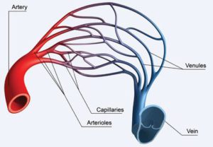
•Movement of fluid requires a pressure (e.g., one created by a pump) to drive it. - The pressure generated by the heart’s beat creates an elevated pressure gradient that pushes blood through to the arteries, then the capillaries, and then back to the veins where it then re-enters the heart. This movement occurs due to the known fact that high-pressure fluids will flow into low-pressure areas.
- Increasing or decreasing the blood flow to areas is controlled by increasing the heartbeat (which allows a faster turnover of fresh oxygenated blood) and constricting arteries or arterioles (small arteries), which reduce or increase blood flow in a specific area.
- 5-6 litres of blood fill the entire circulatory system and continually cycles through the circulation as it is propelled by the heart’s pressure waves.
- Circulation follows the laws of hydrodynamics and hydraulics. By those laws, blood should be evenly distributed throughout the entire fluid circuit of the body.
This model is based off of what is observed in engineered systems where a central mechanical pump is used to push fluid through the system and the resulting fluid motion is then studied. The problem with that model is that what is observed within the body frequently contradicts what is expected according to the model. For example:
- Blood flows are often observed going from low-pressure areas to high-pressure areas (e.g., a capillary bed to veins).
- Blood has been observed to flow spontaneously in the absence of a heartbeat (e.g., after death), and often enters areas with higher pressures on its own.
- Mechanically, the heart provides nowhere near the force that would be needed to push blood through the entire circulatory system.
Note: As mentioned above, I believe liquid crystalline water may be the driving force that makes these inexplicable motions possible. - The pressure, temperature, oxygen saturation and composition of blood, when measured at the same time differ within different portions of the same chamber (ventricle) of the heart. Under the existing models (e.g., simple diffusion or fluid hydraulics), this should not be possible. Note: As discussed in a previous article, I believe liquid crystalline water causes water throughout the body to not mix and instead have different components and constituents from immediately adjacent surrounding water.
- When the blood flow to arteries is measured with rheograms, large arteries will periodically have no blood come to them even though a heartbeat did occur. Note: Riddick also noticed this, but did not look at multiple arteries simultaneously when evaluating pulse pressure waves, and hence assumed no blood was being injected by the heart, when it may instead have simply not been directed to the artery he was measuring.
- The rate of blood flow in the smallest blood vessels (capillaries) within a tissue such as a muscle can rapidly change in a few seconds while no change occurs in the tiny arteries that feed those vessels. Why this happens has been a longstanding question in physiology, and to my knowledge, an answer has not yet been found.
- Likewise, there can be a huge increase in the blood flow to one organ but not the adjacent organ (Goncharenko cited the example of one kidney at times being measured to have 14 times the blood flow of the adjacent one despite the arteries to both remaining at the same diameter). Goncharenko also noted that in cases where there is a systemic total collapse of blood pressure, normal blood pressure will be maintained in the carotid arteries (which is the most important blood supply to maintain since it feeds the brain).
- The total volume of the circulatory system is much greater than the total amount of blood in it. For example, when embalmers drain the blood, 5-6L come out (the total human blood volume), yet when they embalm it, and attempt to fill the vascular system, it often takes 25-30L of the embalming fluid (note: I was unable to assess this claim by Goncharenko). Likewise, when patients are placed on a heart-lung machine (which bypasses these organs while a heart operation is being performed) much more blood (7-15L) is needed than the normal human blood volume.
- Blood can sometimes “shrink.” For example, Goncharenko cited that when embalmers drain blood from the body, 7-8 litres often come out, but when stored, it shrinks to being 6.5 litres. Likewise, he cited examples of 1-2 litres of foreign blood being administered which decreased the total blood volume of a patient rather than increasing it.
- Likewise, instances exist where, according to Goncharenko, the blood volume can expand. This includes when someone begins intense exercise, certain positional changes, the blood volume in marathon runners increasing at the end of a run (despite significant water loss through sweating), and large inexplicable volume changes within the heart (e.g., the left ventricle’s blood volume tripling in size when the heart begins to contract isometrically and valves are closed so no blood can enter it). He also noted that breathing patterns (including shortness of breath), stress and emotions can significantly increase blood volume.
As you consider those points, three possible interpretations emerge.
- Goncharenko’s research was either fraudulent or highly prone to errors.
- Our knowledge of the heart is very primitive and there is much we still need to understand about it (e.g., its blood conjugation).
- Other physical processes are at work (e.g., the vortexes the heart uses to direct blood through the body) that we have almost no knowledge of.
Cavitation Bubbles
One of the most interesting animals in the ocean are mantis shrimps. This is because:
- Their eyes have 16 different colour receptors (compared to just red, yellow, and blue in humans), which allows them to see light polarisation throughout the ocean.
- They have the fastest strike in nature (the limb they use to strike prey can accelerate at the same speed as a 22-calibre bullet being fired).
- That strike has a double impact, which occurs due to the strike creating a cavitation bubble followed by a shockwave which can have almost three times as much force as the initial impact.
In order for water to boil, the energy in it needs to exceed the surrounding pressure which keeps it in a liquid phase. In most cases, we “boil” water by heating it to 100 degrees, and as many mountain adventurers know, water boils at a lower temperature at high altitudes due to less air pressure existing to push it into a liquid phase.
One way to drop the pressure of water is to create a vacuum by expanding the space between two objects faster than the surrounding water can fill it (something which occurs immediately after the mantis shrimp hits its target due to the recoil). This pressure drop causes a cavitation bubble to form which then collapses shortly afterwards once the surrounding water pressure re-enters that cavity. Since this takes place very quickly, the bubble which forms after a mantis shrimp strike only becomes possible to see as a single frame on cameras which capture approximately 40,000 frames per second.
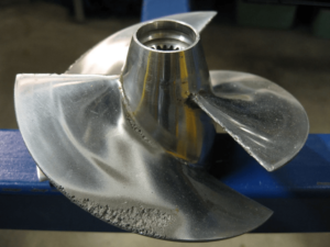
The formation and collapse of cavitation bubbles releases significant amounts of energy both as light and heat and the mantis shrimp cavitation bubble has points that are estimated to be significantly hotter than the sun. The shockwave that follows the formation of cavitation bubbles is significant and is a regular consideration in engineering because if a vacuum is created in water by the motion of a machine, it will gradually destroy that machine. For example, look at the damage done to this steel propeller by the cavitation bubbles it created while pushing a boat forward.
Goncharenko’s team in turn was stuck with answering three seemingly impossible questions:
- How does the propulsive force exist that directs blood to target areas throughout the arterial system?
- How can blood rapidly increase or decrease its volume?
- How is the body able to make do with much less blood than is necessary to fill the entire circulatory system?
The path to a solution we saw in the phenomena occurring with blood in the heart-lung machine. When blood is pumped from veins [into the machine] bubbles appear in it, it foams and increases in volume.
Note: One of the more intriguing therapies I have come across for treating a variety of things, including covid vaccine injuries, involves filtering the blood and exposing it to an oxidative therapy. After each session, a foam is found on the filter (mirroring Goncharenko’s observation) and when examined under the microscope, the foam typically contains a significant amount of microplastics (something many are concerned has become a significant health issue).
Goncharenko thus hypothesised that cavitation bubbles regularly formed in the blood (which contains a significant degree of dissolved gasses to begin with) and that a key function of the heart was to guide the formation of these bubbles. He then gathered pieces of evidence to support his theory including:
- Haematocrit measures how much of blood is composed of blood cells. As you move further away from the heart (especially once you enter veins), haematocrit increases. As far as I can tell, the only way this could be possible is if the blood is initially being diluted by gas bubbles which then leave it.
- Flash-frozen blood contains microbubbles.
- Blood taken from the body and placed into tubes moved spontaneously and microbubbles could be seen within it directing the motion.
Note: I could also see this motion coming from liquid crystalline water. - Cavitation bubbles release sound, light, and electricity. Sound and electricity are detected during a heartbeat but are typically assumed to be from other sources. In subsequent experiments, it was shown that cavitations within blood created electric and sound impulses matching those observed with each heartbeat along with bursts of light in the blood and the blood being pushed along by the cavitations.
Note: Many spiritual traditions express that the body is full of light, something which to some extent has been validated by ultra-sensitive photodetectors that detect photons emitted from cells which appear to regulate many important facets of biology. A century ago, researchers discovered biological photons emitted from cells that caused cells to thrive and multiply (by dividing) – an amazing but largely forgotten side of medicine. One of their many discoveries was that healthy blood emitted the photons (which may be why the heart preferentially sends it to the brain). I mention all of this because I suspect the light generated by the cavitation bubbles may be part of where this property of blood comes from.
The heart appeared to meet the theoretical needs for creating cavitations (it expands, undergoes isometric contractions, and has cross-currents of blood entering it) so imaging was collected to substantiate this hypothesis:
Contrast-enhanced Doppler echocardiography registered the appearance of voids (cavities) in the amount of blood in heart cavities in the brief moment when the regurgitated jet of blood was leaving it. The appearance of caverns in the cavities of the heart coincides in time with the short-term reduction in blood volume and pressure drop in it.
Goncharenko’s team then created some simplified models which showed similar conditions to those inside the heart that would create cavitation bubbles. Most importantly, they observed that while cavitation bubbles were created within water (which expanded by 0.5-1.5% in volume) when blood was placed within the simulated heart, it expanded by 12-22%, indicating that blood is specifically suited for creating cavitation bubbles. Likewise, venous blood had a greater expansion than arterial blood, which was consistent with the previous observation venous blood was more tightly packed than arterial blood (since it has smaller and fewer cavitation bubbles within it).
Assuming his theory is correct, this provides a mechanism to explain how blood could rapidly expand in volume as needed (as it effectively expands like foam) and provides a potential energy source to propel blood through circulation (as the formation and collapse of cavitation bubbles releases significant amounts of energy – something Goncharenko argued was the primary source of the heart’s pumping power).
One of the aspects I find the most intriguing about this entire theory is that within Chinese Medicine, there is a belief that the lungs are responsible for moving the blood through the body, and a variety of breathing exercises exist that seem to do just that when you try them out. Why this works never made sense to me and Goncharenko’s model provides a very elegant explanation.
Conclusion
One of the aspects that continually amazes me is how much people with relatively primitive instrumentation were able to figure out about the body. In the case of the research put forward here, much of it was done over fifty years ago (something that was likewise the case for many other areas I’ve previously covered like blood sludging and zeta potential).
This to me speaks of the issue with modern research – that science is no longer producing paradigm-transforming discoveries, and when independent scientists nonetheless make them, the orthodox scientific community typically bands together to denounce those discoveries. In short, because there is so much money in science, science in the wealthier nations has become a career where the goal is protecting one’s career, not advancing science. If elements like this could be discovered with instrumentation from half a century ago, imagine for a second what our modern scientific apparatus could do if scientists were incentivised to pursue unorthodox research.
Presently, I believe Goncharenko’s thesis of conjugated heart ties is valid, but I am less sure about the other aspects (e.g., the cavitation bubbles) since they will require an independent and unbiased corroboration – something unlikely to be found in the current era. That said, if we simply assume the heart-arterial conjugations are true, this completely changes many of the beliefs that underlie medical practice. For example, it helps to explain:
- Why it has not been possible to make a mechanical pump that effectively replaces the heart – making an artificial heart that can replicate blood sorting, conjugation and vortexing borders on impossible.
- What causes heart attacks and circulatory diseases (e.g., some of my colleagues who have success treating in complex medical issues essentially do so by working through or with the heart-arterial conjugations.
- How the heart has a consciousness and is connected to the entire body (something many different traditions believe).
- How the body solved the problem of not having enough space for all of its necessary blood vessels. Space is one of the key limiting factors in biology and as a, result the human body is very tightly packed with everything needed to support life. So, by allowing the heart to direct both the volume and distribution of blood, it radically increases the space for other essential tissues.
- Why arteries (but not veins) are vulnerable to endothelial damage which then leads to heart disease – as the shockwave from each cavitation the heart creates can be quite powerful and might damage the endothelium if it was sent out on a vector that causes it to collide with the arteries rather than smoothly transit through them.
Note: The above bullet points are a fraction of the potential implications of this research and since there are so many, it is simply not possible to list all of them.
One of my core beliefs is that if something is true, different systems will inevitably rediscover it. Consider for example in Chinese Medicine that the heart is viewed as the emperor that coordinates the functioning of the entire body, something that initially seems implausible. Yet this begins to make much more sense if the heart in fact is responsible for watching everything in the body, sorting what blood goes to each tissue, protects the entire arterial system from damage, and emits a repeating electrical signal that entrains the tissues of the body. I feel this is particularly important because some of the most useful medical approaches I’ve come across for treating complex illnesses appear to operate through utilising these capacities of the heart.
Furthermore, in Chinese Medicine, the heart is viewed as the “fire element” organ of the body, and a fundamental characteristic of “fire” is that it travels in a spiralling pattern. This seems abstract until you realise that that is exactly how the heart moves blood through the body.
This was a lot of ground to cover and I thank you from the bottom of my heart for sticking through all of it with an open consciousness.
Author: A Midwestern Doctor
yogaesoteric
October 6, 2023