Study: MMR vaccine linked to type 1 diabetes

Bioinformatics analysis links type 1 diabetes to vaccines contaminated with animal proteins and autoreactive T cells express skin homing receptors consistent with injected vaccines as causal agent.
Background
Autoimmunity against glutamate decarboxylase (GAD) has been associated with type 1 diabetes. GAD65 (65kDa) and GAD67 (67kDa, to a lesser extent) are involved in type 1 diabetes.
Following an anecdotal report of type 1 diabetes diagnosis a few weeks after measles, mumps and rubella (MMR) vaccine administration, MMR vaccine contents were examined. Major proteins in the vaccine apart from the measles, mumps and rubella live viruses were chicken embryo cell culture proteins.
GAD65 and GAD67 are expressed during chicken embryogenesis.
Method
GAD65 Gallus gallus (chicken) protein sequence was obtained from Uniprot.
BLASTP was used to determine homology to human GAD65.
Results
GAD65 protein comparison between human and chicken reveals 95% sequence homology.
Discussion
The results above provide strong evidence that chicken embryo cell culture proteins in the MMR vaccine can cause the development of antibodies against chicken GAD65 which cross-react with human GAD65 protein to cause type 1 diabetes.
This is very similar to the Pandemrix vaccine causing narcolepsy.
Action
Vaccine design and safety processes have fundamental problems that need to be immediately addressed to avoid such devastating consequences.
Read the full study here.
Vaccines are contaminated with GAD65 (glutamic acid decarboxylase 65 KDa) and AQP4 (aquaporin-4) protein containing chick embryo cell cultures. The role of such contamination in the etiology of type 1 diabetes (T1D) and neuromyelitis optica spectrum disorders (NMOSD), was previously described.
Origin of autoreactive T cells Thymocytes with T cell receptors (TCR) that have high affinity for self peptides are usually negatively selected and destroyed in the thymus. T cells with TCR that recognize peptides that differ by as little as one amino acid from a self peptide, can be positively selected and migrate to the periphery. These low affinity self reactive (LASR) T cells can become autoreactive when they are activated by a peptide that exactly matches a self peptide except for this one amino acid difference (cognate peptide). LASR T cells have the highest possible affinity to self peptides that still qualifies for positive selection in the thymus. This arrangement seems to have worked very well over millions of years of evolution, until human ingenuity intervened…
Vertebrate animal proteins are very similar to human proteins. Animal protein sequences exactly match human proteins except for occasional amino acid differences. So animal proteins are an ideal source of peptides to activate such LASR T cells described above. Ingested animal proteins are immunologically processed to produce a tolerogenic response. Injected animal proteins are however another matter. Animal cells and proteins are used as growth media for the culture of viruses and bacteria, and as excipients in the manufacture of vaccines. Such vaccines are contaminated with numerous animal proteins. When animal proteins are injected with vaccines that either contain live viruses or adjuvants that provoke an immune response, LASR T cells can be activated by peptides derived from these animal proteins thus resulting in autoimmunity.
Immunotoxic effects of injected proteins have been known for over a hundred years. But ignorant vaccine regulators have placed no controls on the proteins that contaminate vaccines. The outcome was predictable. The Pandemrix vaccine was contaminated with influenza virus nucleoproteins which resulted in narcolepsy. Rabbit and duck embryo neural tissue contaminated rabies vaccines cause “neuroparalytic accidents”. Bovine casein and casamino acids used in vaccines are derived from cow’s milk. Therefore cow’s milk proteins contaminate numerous vaccines, contributing to various disorders.
Normally, LASR T cells do not encounter their cognate peptides. Also, due to the lack of innate immune system-derived costimulation, LASR T cells will not be activated if they encounter self peptides complexed with major histocompatibility complex (MHC) molecules on the surface of antigen-presenting cells (APC). Following vaccine administration however, innate immune system costimulation exists. Both live virus vaccines and aluminum adjuvanted subunit vaccines activate the innate immune system. Animal protein derived peptides from the vaccines are complexed with MHC on the surface of APCs and presented to LASR T cells. This combination of innate immune system costimulation and LASR T cells encountering their cognate peptides on the surface of APCs, can result in LASR T cell activation and abrogation of peripheral tolerance.
During evolution, animal protein injection into humans was a rare event. Therefore our immune systems did not have an opportunity to evolve a defense for this mode of abuse.
Determining animal proteins responsible for activation of autoreactive T cells
Various vaccines are contaminated with cow, pig, African green monkey, guinea pig and chick embryo proteins. Homology of these animal antigens to epitopes associated with T1D:
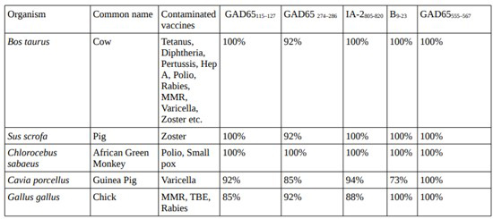
T1D epitope alignment to regions of near identity
Examining the GAD65115–127 and GAD65 274–286 epitopes associated with type 1 diabetes, it is clear that the epitopes are from locations where the chick GAD65 residues differ slightly from the human GAD65 protein (highlighted in figure below). This is exactly what we would expect in LASR T cell mediated autoimmunity. Such chick GAD65 epitopes can activate LASR T cells. Such activated LASR T cells can cross react with human GAD65 and destroy beta cells. Such autoreactive T cells are functional even with low affinity or very low affinity binding. Peptides from locations where the homology between chick and human proteins is 100% are less likely to activate T cells because such T cells are likely to have been negatively selected in the thymus.
Uniprot and BLASTP are used to perform protein sequence alignment analysis.
Homing marker provides evidence of T cell activation site
Hedman et al. write: “No significant differences were seen in any of the Th1-associated receptors, and only the percentages of CD8+ cells expressing the Th2-associated receptor CCR4 was higher in the diabetic group in comparison with healthy individuals, but not observed on CD4+ cells.”
As Berin et al. describe, the CCR4 receptor indicates skin homing and the site of initial priming. Skin homing would be expected since the animal antigen containing vaccines were administered via the subcutaneous or intramuscular route. Such CD8 T cells expressing CCR4 are involved in the destruction of beta cells.
Vaccines disable at least one layer of natural protection against autoimmunity
T cells with high affinity to self peptides being negatively selected in the thymus is one major layer of natural defense against autoimmunity.
T cells in the periphery need to be activated with a self peptide. This is another major layer of natural defense against autoimmunity.
Vaccines are contaminated with numerous animal proteins. Animal proteins contain numerous peptides identical to self peptides. Upon administration of such vaccines, with innate immune system-derived costimulation active, these animal protein derived peptides activate T cells that have escaped thymic negative selection. So this layer of defense is disabled.
Protein sequence alignment
Here we analyze protein sequence alignment between T1D autoantigens and various viral, bacterial, fungal proteins that contaminate vaccines along with animal proteins. Chick and Coxsackievirus protein sequence alignment scores are also included for comparison. Natural infection with Coxsackievirus has been linked to T1D.
Method
BLASTP methodology was used for protein sequence alignment. As shown before, a BLASTP sequence alignment score of 19.3 was obtained comparing human hypocretin receptor and H1N1 nucleoprotein contained in the Pandemrix vaccine. This level of sequence alignment was sufficient to cause autoimmunity that resulted in hypocretin dysregulation and narcolepsy. Therefore any score equal to or higher than 19.3 suggests high probability of autoimmunity.
While vaccines target one or a few particular viral, bacterial proteins, most vaccines are contaminated with all proteins from that virus or bacteria. Example: the Pandemrix vaccine contained both H1N1 hemagglutinin (target) and H1N1 nucleoproteins (contaminant). The exceptions are recombinant vaccines. In recombinant vaccines, the vaccine contains only the target protein from the target organism. The target protein is produced usually by genetically modifying yeast (Saccharomyces cerevisiae). Hepatitis B and HPV vaccines are produced using this technique. Such vaccines are however, contaminated with all Saccharomyces cerevisiae proteins.
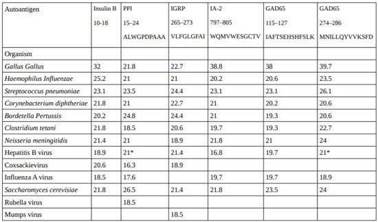
As we can see above, there are numerous scores above the 19.3 baseline that corresponds to the level of sequence alignment in Pandemrix vaccine induced narcolepsy. So these contaminants can contribute to autoimmune diseases.
Conclusion
The above findings add to the growing evidence of vaccines inducing autoimmune diseases.
While the analysis above focused on T1D, the mechanisms described are obviously likely to be active in any number of autoimmune diseases.
It is quite obvious that there are fundamental problems with vaccine design and safety. Vaccine designers need to go back to the drawing board. We need vaccines that are safe by design.
yogaesoteric
December 14, 2018